

Today is our topic of discussion Definition of Strongyloidiasis
Definition of Strongyloidiasis
Definition:
Infection with Strongyloid stercoralis is called ‘strongyloidiasis’.
Geographical distribution:
World wide. Common in Brazil, Cambodia, Philippines and Africa.
Epidemiological determinants:
Agent:
Strongyloid stercoralis
Egg:
Laid within mucous membrane from where larvae hatches out and comes to the lumen of intestine from where passes outside with stool. So, only larvae are seen not the eggs.


Larvae:
Two types-
1. Rhabditiform larva (1st stage larva): Developed directly from gravid female and found in lumen of bowel.
2. Filariform larvae (Infective larva): Is the infective form. Enter body of human through skin. Host: Man – Live in the tunnel of mucous membrane of small intestine
Mode of transmission:
1. Infective larvae in infected soil penetrate human skin.
2. Person-to-person transmission is rare, but has been documented.
3. Travelers visiting endemic areas and have contact with contaminated soil through bare skin are at risk.
Life cycle:
First stage larvae pass out in feces → Develop in faeces on ground to infective larvae or free-living adults mate & females lay eggs which hatch → Develop into infective larvae → Penetrate the skin on contact with soil → Enter superficial veins and then blood vessels to alveoli of lungs → Coughed up and swallowed → Parasitize the intestinal mucosa (duodenum and jejunum) → Only females will reach reproductive adulthood in the intestine.
Female strongyloides reproduce through parthenogenesis (eggs hatch in the mucosa of intestine) → Young larvae excreted in feces (so, S. stercoralis causes both respiratory and gastrointestinal symptoms) or development of 1st stage larva into infective larvae in the gut of the host by autoinfection → Autoinfective larvae penetrates wall of lower ileum or colon or skin of perianal region →
Enters circulation again and to lungs→ Back down to the small intestine thus repeating the cycle. The free-living males and females of S. stercoralis die after one generation; they do not persist in the soil.
[Autoinfection makes strongyloidiasis a life-long infection unless effective treatment eliminates all adult parasites and migrating autoinfective larvae]
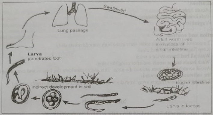

Fig: Life cycle of Strongyloides stercoralis
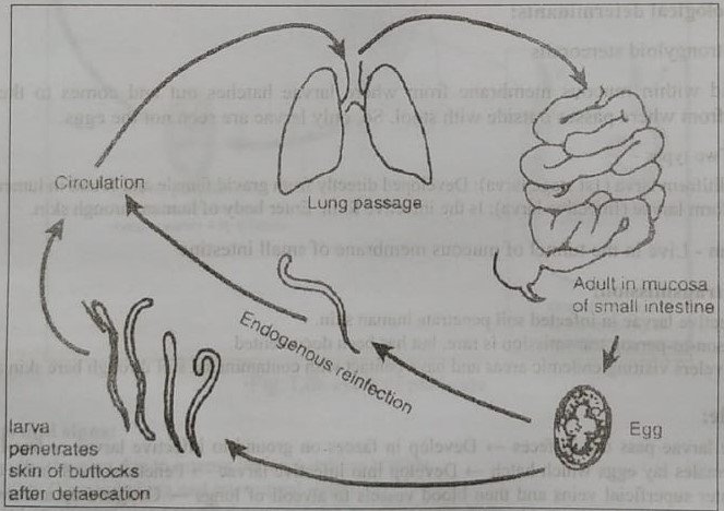

Fig: Mode of autoinfection with Strongyloides stercoralis
Signs and symptoms:
Dermatitis:
Swelling and itching at the site where the skin has been penetrated.
Lungs:
Wheezing and coughing, along with pneumonia-like symptoms (Loeffler’s syndrome).
Intestine:
Burning pain, ulcer, diarrhoea, vomitting & in severe cases, intestinal obstruction
Weight loss.
Immunocompromised states or individuals taking corticosteroids:
Cause a hyperinfective syndrome (or disseminated strongyloidiasis) due to the reproductive capacity of the parasite inside the host.
Diagnosis:
1. Microscopic examination: Demonstration of larvae in stool sample.
2. Demonstration of larva in sputum.
3. Blood: Eosinophilia
4. Serodiagnosis by ELISA.
Complications:
1. Acute pulmonary eosinophilia (Loeffler’s syndrome).
2. Disseminated strongyloidiasis: Especially in patients with HIV or in compromised immunity system.
3. Malnutrition due to malabsorption from GIT.
Treatment:
Ivermectin, Albendazole and Mebendazole (dose in previous pages
Control and preventive measures:
1. Treatment of the cases & regular antihelminthic prophylaxis.
2. Improved sanitation system & proper disposal of feces.
3. Practicing good hygiene (washing of hands), etc.
See also :